The Critical Foot-Brain Connection
A Guide to Natural Gait Mechanics and Therapeutic Foot Care
Lesson 2 – Optimal Foot and Lower Limb Function
If the video player does not load, please ensure your browser allows embedded video.
Downloads: Lesson 2 Course Book / Monograph
The Gait Cycle
(For more detailed coverage of this topic, refer to The Future of Foot Care Monograph, pages 5- 26.)
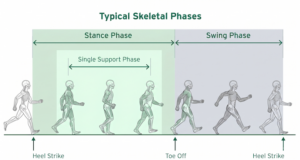
Figure 2.1. Typical skeletal phases.
Optimal Neuromusculoskeletal Mechanics During Gait
Optimal neuromusculoskeletal mechanics are typically and exclusively observed within individuals who are in a habitual barefoot environment. When barefoot, the sole of the foot picks up the subtle variations in terrain (texture and orientation) and this tactile stimulus from the ground is not dampened. The brain uses these tactile stimuli, in concert with the proprioceptive stimuli received from the feet, ankles, legs, hips, and back to initiate protective muscle activations throughout the lower limbs such that they are capable of safely managing the dynamic forces generated by the demands of three-dimensional activities. When barefoot, the foot is unfettered and thus there is no restriction to the dynamic musculoskeletal movement. When barefoot, the foot receives Right Stimulus and, as a result, Right Movement is uninhibited.
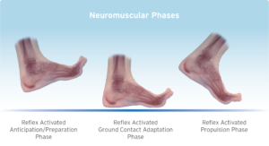
Figure 2.2. Neuromuscular phases.
Neuromuscular Reflex-Activated Anticipation/Preparation Phase
Movement-generated proprioceptive sensory input and tactile stimulus (from the foot’s ground contact) initiates a protective reflex response during the skeletal swing phase (while the foot is off the ground) in anticipation of the imminent next step’s ground contact.
Neuromuscular Reflex-Activated Ground Contact and Propulsion Phases
The protective reflex response activates muscle contractions to optimally align and stabilize the bones of the foot, ankle, leg, and hip in order to safely manage the forces anticipated during the next step’s ground contact (skeletal heel stance phase). The resultant optimal alignment and stability not only protects the structure, but also ensures optimal muscular efficiencies and performance capabilities.
Activity-related stresses are safely managed and, therefore, contribute to the neuromusculoskeletal structure becoming more robust (i.e., stronger and more flexible), thus significantly reducing the risk of injury.
Ideal Gait Mechanics of the Foot and the “Optimal Arch Apex”
During natural healthy foot function (as observed in the habitually unshod community), optimal neuromuscular function and related musculoskeletal mechanics/alignment are ideally a dynamic response to activity levels and terrain. In other words, the muscles of the foot should act to optimally align the bones to most effectively manage the forces generated during varying activities and terrain. Thus, the dynamic stable arch system would provide a capable foundation for the kinetic chain of the lower limbs and body while promoting optimal neuromusculoskeletal alignment/function/performance so that little or no degenerative stress is generated throughout the kinetic chain.
From a strictly mechanical perspective, the lower limb structure can be considered to comprise a ball and socket joint at the hip, a simple hinge joint at the knee, with the foot and ankle functioning similar to that of one- half of a universal joint, in order to provide an effective interface with the ground. However, closer examination of the skeletal structure of the foot and ankle suggests that, with appropriate muscle contractions, the bones of the foot are capable of aligning into a dome-like configuration, which can thus behave much like a socket moving around an imaginary ball. (Figure 2.3).
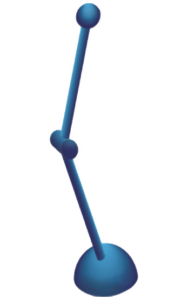 Figure 2.3.
To date, it has been widely accepted that the shape of the interlocking bones and ligament strength combine to maintain the transverse, medial longitudinal, and lateral longitudinal arches of the foot. This established viewpoint, while technically correct, overstates the role that bone shape and ligament strength play in maintaining optimal structural integrity of the foot. For example, if we isolate the bones of the foot from the muscle, tendons, ligaments, etc., and view the structure from a physics perspective, it becomes clear that the relative alignment and positioning of the bones are the primary determining factors in the foot’s structural capabilities.
Within the medical community, the foot is commonly described as consisting of the medial longitudinal, lateral longitudinal, and transverse arches. This view, from a physics perspective, is inordinately simplified and ignores the complexity of the structure as a whole. The structural physics of the foot more accurately demonstrates a series of intersecting arches that run medially to laterally and posteriorly to anteriorly from the calcaneus to the metatarsal heads. To better understand both the simplicity and complexity of this arch system, it is important to identify the dynamics of a single arch and its intrinsic relationship within a system of arches.
In the foot, the structural mechanics of a single arch (Figure 2.4) are determined by its components:
- The material composition of the arch: interlocking bone structure and ligaments – their relative strengths (e.g., tensile, compressive.) and elasticity.
- A tie beam: soft tissue, i.e., tendons, muscles, fascia, etc., and their relative strengths (tensile and elastic).
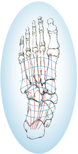
Figure 2.4. The multitude of individual arches that form the foot’s arch system.
Within the material composition of any given arch structure, there exists a central “keystone” about which opposing forces must equalize as a means of maintaining the arch integrity. When force is applied to an arch structure, the stronger and more stable the material composition, the lower the degree of tensile (or pulling) force produced on the tie beam.
When combined in a multi-arch system such as the foot, these singular arch dynamics work synergistically to maximize relative strength and stability while greatly minimizing stress, and are more effective collectively than individually.
Therefore, from a physics perspective, the most inherently sound structural mechanics would be achieved if the bones of the foot could interlock and maintain the multi-arch functional dynamics of a dome shape. Such a dynamic could manage greater loads with minimal contribution from, or stress on, the ligaments and extrinsic/ intrinsic musculature. The dome shape of the interlocking bones would function much like a socket, capable of rotating around an imaginary ball (Figures 2.3 and 2.5). The level of functional stability of the dome would be determined by the “ideal” or “optimal arch apex” height necessary to most effectively maintain structural integrity in the interlocking bones as they manage the forces generated throughout three-dimensional activity.
Further, the location of the “optimal arch apex” would ideally correlate to the location of the “conceptual” arch keystone for optimal force management.
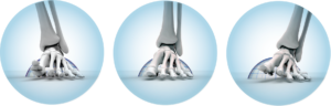
Figure 2.5. The dome shape of the interlocking bones.
The relative positioning of the mid- foot joints (i.e., the optimal arch apex) is significant to the degree and pattern of forefoot segment motion, which in turn, is indicative of the foot’s stability.
Biomechanically, and within the kinetic chain model, the integrity of the foot’s structural alignment plays a significant role in managing the forces and stresses generated during gait. It is clear that an ideal dome-like structural alignment in the foot is possible, and that there is an inverse relationship between the structural integrity of the foot and the muscular effort required to facilitate and manage its relative alignment. The more structurally sound the arch, the less muscular effort is required to manage the alignment.
As indicated earlier, during ideal natural healthy foot function, the optimal neuromuscular function and related skeletal alignment is a dynamic (protective) reflex response to the activity levels and terrain currently being experienced. That is, the muscles of the foot act to optimally align the bones to most effectively manage the forces generated during varying activities and terrain prior to each step’s ground contact. For example, while running, tactile, nociceptive, mechanoreceptive and proprioceptive stimuli trigger reflex muscle activations to proactively create a higher (mechanically stronger) and more stable arch system than when walking. Thus, the protective-reflex based and activated, dynamic, stable arch system provides a capable foundation for the lower limbs and body (kinetic chain) while promoting optimal neuromusculoskeletal alignment/function and minimal degenerative stress throughout.
An excellent example of neuromusculoskeletal conditioning potential can be found in individuals who have lost their arms, yet developed the dexterity of their feet to the extent that they function as “hands” – still capable of performing many complex tasks, all with a considerable degree of finesse and precision. Extrapolating from this model, there is no reason that the neuromusculoskeletal function of the feet cannot be conditioned to achieve ideal, dynamically domed, structural alignment, as described in the previous Lesson.
It would be virtually impossible to quantify the role of specific muscles throughout such a multiplicity of activities. We can, however, examine the relative roles (primary and supporting) that muscles are ideally capable of performing throughout the gait cycle, from a mechanical perspective.
The extrinsic muscles of the foot comprise the extensors (originating in the lateral aspect of the shin), the flexors (originating in the posterior side of the lower leg) – both groups are connected to the foot via long tendons – and the ankle flexors (i.e., the calf muscles). The intrinsic muscles of the foot (located primarily in the plantar region of the foot) comprise flexors, adductors, and abductors.
From an ideal mechanical perspective, the following muscles are grouped according to their gait-related roles (see Figures 2.6 and 2.7):
Prior to weight bearing:
- Alignment of the foot and ankle structure: via active extrinsics — extensor hallucis longus and digitorum longus, tibialis anterior, and peroneus longus (aka fibularis longus) (Group A).
- Stabilization of the foot and ankle structure: via active Group A (re: foot), Peroneus longus active peroneus brevis (aka fibularis brevis) and tibialis posterior (Group B) (re: ankle), in concert with passive extrinsics – flexors hallucis longus and brevis and digitorum longus (Group C), and passive intrinsics – quadratus plantae and flexors digitorum brevis & minimus, and lumbricals (Group D).
During weight bearing:
- Stabilization of the foot and ankle structure: via active Group A and peroneus brevis (aka fibularis brevis) with passive to active Group B, Group C, and abductors hallucis and digitorum minimi, adductor hallucis and the interossei (Group E), in addition to the plantar fascia (Group F).
Propulsion:
- Stabilization of the foot structure: via active Groups B, C, D, and E and Tibialis posterior (origin) active to passive Group A and peroneus brevis (aka fibularis brevis).
- Propulsion: via active Group B and active extrinsics – gastrocnemius and soleus.
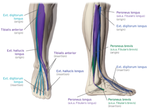
Figure 2.6.
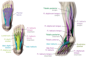
Figure 2.7
Ideal Neuromuscular Gait Mechanics — The Windlass and Cuboid Pulley Effects
If the supporting musculature of the foot aligned and stabilized its interlocking bones into a functionally dynamic dome shape prior to weight bearing, the structure would be inherently strong and resilient. This would provide the most stable and stress-free foundation for the rest of the body, requiring the lowest degree of muscular effort during the weight bearing and propulsion phases of gait.
This alignment and stabilization process is, indeed, exhibited in barefoot gait, and is easily achieved during the swing phase as the foot moves from the muscle-firing sequences of propulsion to the extensor muscle-firing sequences of dorsiflexion (Figures 2.8 and 2.9).
When examining the muscle-firing sequences of the lower leg extensors During the gait cycle, EMG analysis shows a co- contraction of the peroneus longus (a.k.a. fibularis longus) and tibialis anterior, prior to heel strike, reinforcing their implied significance in the alignment and stabilization process of the foot and ankle, as discussed in the above section on the kinetic chain.
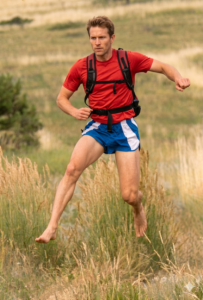 Figure 2.8.
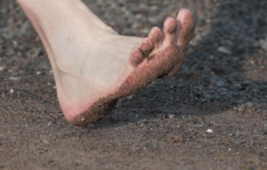 Figure 2.9.
Coupling this information with their respective origins and insertions, these opposing contractions cause a transverse pulling or cinching action that essentially aligns the bones of the foot’s midtarsal region into a dome-like position with an ideal (maximum) transverse arch apex height (Figure 2.10).
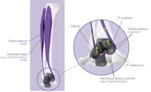
Figure 2.10.
This is further supported by the fact that the main actions of the tibialis anterior are dorsiflexion and inversion, while the main actions of the peroneus longus (aka fibularis longus) are dorsiflexion and eversion. When these two muscles act in functional balance, they have a “stirrup-like” effect as the ground contact base of the posterior fascial line.
The cinching action of the peroneus longus (a.k.a. fibularis longus) tendon around the cuboid is essential to the control of the transverse arch’s feature of stability with adaptability. This process, often called the “Cuboid Pulley Effect,” with the antagonistic activity of the tibialis anterior, establishes the 1st metatarsal/1st cuneiform joint not only as the “conceptual” transverse arch keystone, but as the foundation of the entire kinetic chain, regardless of activity levels and terrain.
The function of the interosseous muscles (i.e., adduction of the 3rd to 5th toes toward the 2nd toe, and abduction of the 2nd to 4th toes) establishes the 2nd ray as the longitudinal axis of the foot’s dome-like functional configuration.
Another important contribution to the dome-like alignment and ideal longitudinal arch apex in the pre-contact phase, is contraction of the extensor hallucis longus; this results in the Windlass Effect of the great toe and plantar flexion of the first metatarsal (Figure 2.10). In addition, simultaneous contraction of the extensor digitorum longus causes dorsiflexion of the corresponding digits and plantar flexion of the related metatarsals. The Windlass Effect is further enhanced, regarding the 2nd to 5th digits, by passive to active tension within the lumbricals, which (via their dorsal insertion points) also contribute to dorsiflexion of the interphalangeal joints.
Of great significance, with a synergistic effect toward optimal arch apex formation, is the role played by the hallux and the sesamoid bones. Together they stabilize and lock into place the Windlass Effect through the 1st ray. Prior to ground contact, simultaneous to the Windlass and Cuboid Pulley activations, the hallux dorsiflexes and the sesamoids glide forward up and distal to the 1st metatarsal head, thus maximizing the tension on the flexor hallucis longus. Once the 1st metatarsal becomes weight bearing, the sesamoids’ location (distal to the 1st metatarsal head) is such that the Windlass Effect is locked in place. This mechanism assures that the optimal arch apex cannot collapse during the entire stance phase of gait (Figure 2.11).
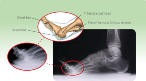
Figure 2.11.
As the digits dorsiflex, the mechanical dynamic that causes plantar-flexion of the metatarsals corresponds to a passive tension or preloading of the following:
- The tendons of flexors hallucis longus (and slip) and digitorum longus, muscle body of quadratus plantae and the lumbricals – the sAbductor hallucis, flexor digitorum brevis, and abductor digiti minimi – the intrinsic first layer muscles, and
- the plantar fascia.
The opposing active tension created between the extrinsic extensors and 1st and 2nd layer muscles cinches the interlocking bones into a dynamic dome-like structure that is capable of handling enormous force with minimal muscular contribution (Figure 2.12). The preloaded intrinsic 1st and 2nd layer muscles and plantar fascia provide a resilient tie beam of optimal tensile strength.
As already described, natural healthy foot function and ideal gait mechanics should demonstrate optimal neuromusculoskeletal function (timing of muscle firing and alignment) throughout the kinetic chain as a dynamic response to activity levels and terrain. That is, subtle variable stimuli to the sole of the foot Optimal “Natural” Foot and Lower Limb Function produces:
- Tactile and nociceptive reflex activations of the foot and ankle related muscles.
- Proprioceptive reflex activations in the muscles throughout the lower limb, hip, and back kinetic chain.
Together, they optimally align the bones to most effectively manage the forces generated during varying activities and terrain while promoting optimal neuromusculoskeletal function and little or no degenerative stress. Tactile, nociceptive, mechanoreceptive, and proprioceptive sensory stimuli of the first step and/or optimal proprioceptive conditioning triggers a protective reflex response during the swing phase of gait prior to the second step ground contact. This continuous, step-by-step, nociceptive/mechanoreceptive/proprioceptive reflex activity results in a pre-ground contact cinching of the interlocking bones of the foot and ankle to:
- Form a strong yet adaptable dome-like shape in the foot (i.e., Optimal Arch Apex [OAA]).
- Lock the foot and ankle to inhibit eversion or inversion at ground contact (i.e., stabilize the subtalar region for optimal mechanical positioning through the knee and hip in line with the arch apex).
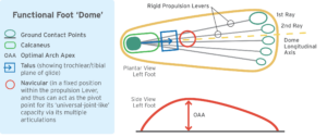 Figure 2.12.
The reflexive pre-ground contact musculoskeletal cinching is a dynamic response to activity levels and terrain. Functioning in this ideal manner, the foot’s neuromusculoskeletal structure is capable of providing optimal structural integrity, alignment, and shock management throughout multidirectional ground contact, weight-bearing, and toe off, while forming a spring-loaded rigid lever when in the propulsion mode.
When the ankle is locked against eversion and inversion at heel contact, the roundness of the heel initiates a smooth, stress-free transition, naturally aligning the forefoot to the ground. This is consistent in multidirectional activity through varying angles of impact.
In short, to paraphrase sports training concepts, the barefoot environment promotes Proper Technique toward the creation of optimal foot function.
Over time, with repetition, the body adapts to the diversified stimuli and optimal neuromusculoskeletal mechanics and ultimately becomes the conditioned norm or “optimal reflexive condition.” This can be “reconditioned” or “retrained” to become a maladapted reflexive condition through Poor Technique activities of sufficient intensity and duration.
Lesson 2 Quiz
Select all answers that apply. A score of 70% or higher is required to pass.
No email is required to take the quizzes. After passing all four lessons in this browser, you can request an emailed certificate of completion.
Request Certificate of Completion
Enter your details exactly as you would like them used for the certificate email.
Please confirm the certificate name before submitting. The emailed certificate will use this text.
Continue to Lesson 3